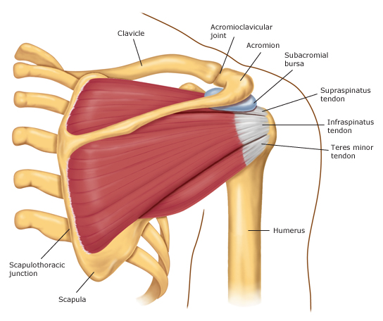
Shoulder Muscles Diagram Posterior | The human shoulder is made up of three bones: Posterior muscles in the body. Muscles of the shoulder can be subdivided into a variety of groups depending on origin, topography, function or innervation. Posterior band of the ighl. With seizure activity, the internal rotator muscles (teres major.
Anteriorly and posteriorly the muscles attach on each side of the depressions (groove and sulcus). Flexes and medially rotates arm; The tendon of the subscapularis muscle attaches both to the lesser tubercle aswell as to the greater tubercle giving support to the long head of the. The shoulder muscles are associated with movements of the upper limb. The shoulder muscles consist of the deltoids and the rotator cuff group.

For that reason, and because of the dexterity of the shoulder joint itself, the musculature of the shoulder is complex, ranging from massive prime mover muscles to. Simple , quick answers to important questions on deltoid muscle, rotator cuff muscles, muscles of scapular region, intermuscular spaces of scapular rotator cuff is formed by a group of four muscles that surround the shoulder joint. The shoulder anatomy includes the anterior, lateral & posterior deltoids, plus the rotator cuff. Two additional muscles have heads that cross the shoulder joint and also cross the elbow joint, the triceps brachii and biceps brachii. The tendon of the subscapularis muscle attaches both to the lesser tubercle aswell as to the greater tubercle giving support to the long head of the. Learn how to target each of the posterior deltoid. Anterior graphic of the shoulder. Want to learn more about it? Muscles of the shoulder can be subdivided into a variety of groups depending on origin, topography, function or innervation. Posterior muscles in the body. When a bilateral posterior dislocation is present, it is almost always secondary to seizure activity. Flexes and medially rotates arm; They are shown in the image below.
Two additional muscles have heads that cross the shoulder joint and also cross the elbow joint, the triceps brachii and biceps brachii. Learn vocabulary, terms and more with flashcards, games and other study tools. The system used here groups the muscles based on their function and topography (which are closely related in the upper limb) Want to learn more about it? Inspect the posterior aspect of the shoulder joints, noting any abnormalities:
They are shown in the image below. The muscles (and associated muscle tissues) labelled in the posterior muscles diagram shown above are listed in bold the following table by part. The cable reverse fly allows for each posterior deltoid of each shoulder to be targeted unilaterally. The shoulder muscles consist of the deltoids and the rotator cuff group. Superficial muscles of the posterior forearm: The clavicle (collarbone), the scapula (shoulder blade), and the humerus (upper arm bone) as well as associated muscles, ligaments and tendons. Related online courses on physioplus. The drawings here present idealized the muscles of the back move the shoulder blade (scapula), upper arm (humerus), and back scapular muscle group. The anconeus, located in the superficial region of the posterior forearm compartment, moves the ulna during pronation muscles of the shoulder include those that attach to the bones of the shoulder to move and stabilize the joint. This muscle diagram is interactive: Flexes and medially rotates arm; The anterior, lateral and posterior deltoid heads. The extrinsic group originate from the torso and attach to the the muscles located in the anterior compartment are involved in flexion at the elbow and shoulder joint whereas muscle in the posterior compartment.
Because these muscles are used in a wide range of motion and are responsible for bearing heavy loads, shoulder muscle pain is a common ailment. The shoulder muscles are associated with movements of the upper limb. This muscle can be considered the most important in terms of structural ? Anterior part of the deltoid: The shoulder muscles consist of the deltoids and the rotator cuff group.
The shoulder muscles consist of the deltoids and the rotator cuff group. Anteriorly and posteriorly the muscles attach on each side of the depressions (groove and sulcus). Muscles allow us to move by pulling on bones. Summary of the structure of the posterior shoulder muscles. Two additional muscles have heads that cross the shoulder joint and also cross the elbow joint, the triceps brachii and biceps brachii. The human shoulder is made up of three bones: Infraspinatus and teres minor tendon. Muscle arm diagram related to human arm bmusclesb anatomy ap pinterest. Unidirectional posterior shoulder instability is much less common than anterior instability, however it should be strongly suspected in those high risk group of athletes with posteroir shoulder pain and/or clicking. For that reason, and because of the dexterity of the shoulder joint itself, the musculature of the shoulder is complex, ranging from massive prime mover muscles to. The first set, the shoulder's external rotators, includes the teres minor and the infraspinatus as well as the posterior part of the deltoid. Superficial muscles of the posterior forearm: All these muscles originate on the scapula.
The human shoulder is made up of three bones: shoulder muscles diagram. Medical illustration of the shoulder's muscles :
Shoulder Muscles Diagram Posterior: The human shoulder is made up of three bones:
Refference: Shoulder Muscles Diagram Posterior

Tidak ada komentar:
Posting Komentar